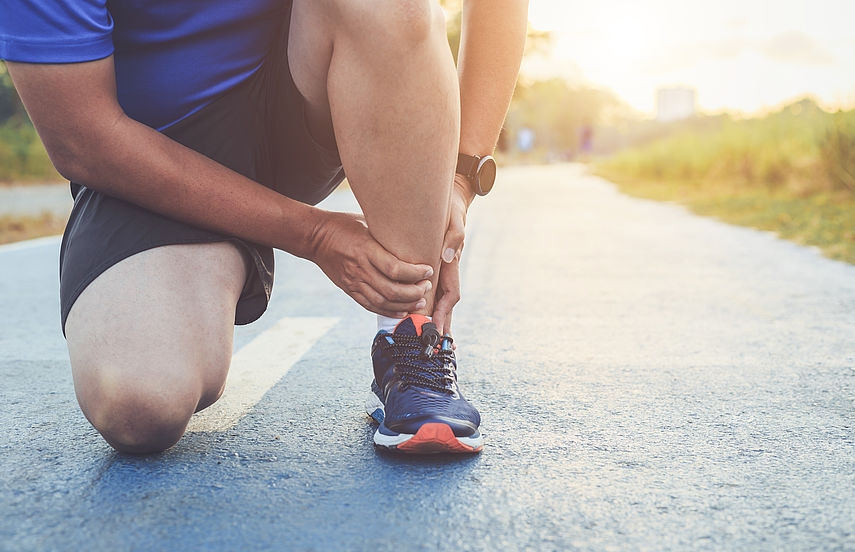
RadNet: A Leader in the Treatment of Sports Injuries

RadNet is heavily involved in sports medicine, particularly in Southern California. Led by Dr. John Crues, head of our sports musculoskeletal program, our radiologists work closely with many of the leading teams in Los Angeles and Orange County, including the hockey team the Anaheim Ducks, the baseball team the L.A. Angels, and the Los Angeles Rams football team. RadNet plays a central role in diagnosing athletes and identifying the probable mechanism behind their injury.
Common Sports Injuries
Though musculoskeletal radiologists work with every part of the human body, the most frequent sports injuries occur in the shoulders, elbows, and knees. Every sport has its own common set of injuries. Shoulder and elbow injuries are more common in baseball, while ligament and cartilage tears are more common in football and basketball, which focus more running and jumping.Over time, these activities can have a deleterious effect on bones, ligaments, and tendons, leading to a common set of tears and fractures.
- Stress Fractures. Stress fractures occur when muscles become tired and lose their ability to absorb impact. When this happens, the stress of impact is transferred to the bones, where it builds up until it causes a tiny crack. Knees are extremely susceptible to stress fractures because they are the largest weight-bearing joints in the body. However, stress fractures can occur in the elbows and shoulders as well, particularly in sports that emphasize arm and shoulder movement, such as pitchers, weight lifters, and rowers.
- ACL Tears. The anterior cruciate ligament (ACL) connects the shinbone to the thigh bone and is one of the most commonly injured ligaments in sports. ACL tears are caused by a direct blow to the knee or repeated stress from jumping, pivoting, improper landings, and sudden stops.
- Ulnar Collateral Ligament Tears. The ulnar collateral ligament (UCL) is located on the inside of the elbow and connects the bones in the upper arm to the bones in the forearm. UCL tears aremost common inbaseball pitchers, however,hockey playersare susceptible to them as well.
- Meniscal Tear. The meniscus is a C-shaped piece of cartilage that cushions and supports the bones in the knee. Tears are caused whenever the knee is twisted too severely or whenever the knee is subjected to repeated stress, such as jumping or sudden stops. Tears are not only painful, they sometimes cause cartilage to break off and lock up the kneejoint.
- Rotator Cuff Tears. The rotator cuff is made up of four tendons that cover the top of the arm bone and are extremely important in sports that involve throwing.The rotator cuff can be torn by a sudden impact, such as falling with the arm outstretched, but tears normally occur after the muscles have been worn down by repeated stress. Baseball pitchers and football quarterbacks are extremely susceptible to these injuries.
Diagnosing Sports Injuries
To diagnose a musculoskeletal injury, RadNet radiologists use a set algorithm to examine every part of the joint that is injured. The algorithm provides a set of injuries for each specific joint while also allowing radiologists to envisage what the healthy joint should look like. By comparing the athlete’s images to the algorithms, RadNet radiologists determine whether an injury is present, how severe it is, and the most probablecause.
Once they’ve finished their diagnosis, the radiologists write a report detailing the athlete’s condition and submit it to the referring doctor. Based on the medical analysis provided by the RadNet team, the referring doctor decides on the appropriate course of treatment. RadNet doctors are also commonly involved in the decision as to whether an athlete should be allowed to continue playing, depending on the severity of their injury.
Imaging Modalities
RadNet typically uses magnetic resonance images (MRI) to diagnose athletes. CT and ultrasound are used as well, but MRI provides better contrast, allowing doctors to examine small structures in greater detail. Over 60 percent of the medical images taken during the 2018 Winter Olympics were taken using MRI. MRI is especially useful when diagnosing bone fractures, because it is far more sensitive to the kinds of subtle injuries that can occur during athletic competition.
RadNet’s musculoskeletal team also participates in various research programs with the sports-medicine group Kerlan-Jobe, as well as The Santa Monica Orthopedic Group (now part of Cedars Sinai Medical Network), helping them develop new surgical techniques to benefit athletes. Additionally, RadNet imaging has helped surgeons better visualize knee cartilage and together, we are running an ongoing study to identify common abnormalities in healthy elbow joints and to perfect the algorithms used to diagnose patients.

Add new comment